Neuro-Imaging Techniques
Neuro imaging techniques
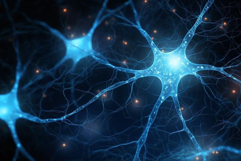
The brain, an organ of the human body that we, as Psychology students, are much interested
in. The brain, weighing about 1.67 kilos for adults, consists of about 60% fat, the other 40% is a combination of water, protein, carbohydrates, and salts. But that is not the only thing it consists of. By taking a look deeper down, it becomes noticeable that the brain also consists of much more, like: blood vessels, neurons, and glial cells (these being the astrocytes, oligodendrocytes, Schwann cells, and microglia). For neuroscience, the focus is of course put on the neurons. But how do we get these active microscopic cells on an image? Well, there are quite a few ways. These methodologies being: EEG, fMRI, MEG, (f)NIRS, PET, TMS, TDCS, and even more. And within this blog, we will go over some of these methodologies.
fMRI
fMRI, also known as functional magnetic resonance imaging, is the same as an MRI but the focus is put on the functioning of the brain during a task. Both fMRI and MRI make use of a large magnet. This magnet, while rotating around the person's head, is able to orient the hydrogen within someone's blood. When this magnet stops emitting radio waves, depending on the amount of oxygen that is within the area, the brain emits a radio wave back to the fMRI machine. Thus, creating an image that showcases activity within the brain. This activity within the brain is able to be showcased, because activity within the brain results in more oxygen within the brain areas that is correlated with that specific activity. Thus, more oxygen equals more brain activity, equals visibility of activity on the fMRI scan.
fNIRS
fNIRS, also known as functional near-infrared spectroscopy, is the same as NIRS, but just like the fMRI, the focus is put on the brain activity. Instead of a magnet that (f)MRI uses, fNIRS makes use of emitted light, as the human body is quite transparent for infrared light. Thus, fNIRS looks at the changes of light absorption within the human body, from which it is possible to calculate
the changes in concentration of oxygenated and deoxygenated blood. As stated previously, whenever activity within the brain occurs more oxygen is required for the action to occur. Thus, when an action occurs, the brain requires more oxygen. This change from deoxygenated to oxygenated blood is able to be measured via near-infrared light, which is able to give us an image of where in the brain activity
is occurring.
EEG
But why add something, when we can also measure something that is already there? That is where the EEG, also known as the electroencephalogram, comes into play. Instead of adding light, or radiowaves, an EEG looks at the electrical currents that are occurring within and between neurons. These currents are able to be picked up via an electrode, which is somewhat able to showcase which parts of the brain are active when an action occurs. Instead of looking at changes of oxygen, the EEG is looking at changes of potassium, sodium, calcium, and chloride within the brain. These four ions are either positively or negatively charged. When a neuron gets activated, its electrical charge goes from -70mV to +40mV. This change in voltage is able to be measured by the EEG.Besides these three, there are still many more to talk about. However, if I had to explain all of them, this blog would turn out to be way too long. Thus, if you are interested in learning more about the brain and/or how to measure its activity. Either think back about our beautiful module 3, or perhaps think about taking a look at the neuroscience minor that you can take in your third year.